Dot-Blot Assay for dsRNA in IVT RNA
Detects double-stranded RNA (dsRNA) contaminants in RNA made by in vitro transcription, such as crRNA transcripts, using a J2 anti-dsRNA primary antibody, an HRP-conjugated secondary antibody and chemiluminescent detection. The J2 antibody recognises dsRNA regions longer than about 40 nucleotides, which makes the dot blot a convenient way to compare dsRNA levels between IVT reactions. Allow 4-6 h.
Contents
Section titled “Contents”- Materials
- Sample and membrane preparation
- Membrane blocking
- Primary antibody incubation
- Secondary antibody incubation
- Chemiluminescent detection
- Imaging and analysis
- Notes
- References
1. Materials
Section titled “1. Materials”1.1 Reagents and supplies
Section titled “1.1 Reagents and supplies”| Reagent | Manufacturer | Catalogue number |
|---|---|---|
| Biodyne nylon membrane | Thermo Fisher Scientific | 77016 |
| J2 anti-dsRNA antibody (mouse) | Sigma-Aldrich | MABE1134-100UL |
| HRP-conjugated anti-mouse secondary antibody | Thermo Fisher Scientific | 31430 |
| Poly(dI) dsRNA control | Sigma-Aldrich | P1530 |
| Non-fat dry milk powder | Carnation | - |
| SuperSignal West Pico PLUS | Thermo Fisher Scientific | 34577 |
| SuperSignal West Femto | Thermo Fisher Scientific | 34095 |
| 10x TBS | - | - |
| 10x TBST | - | - |
| RNase-free water | - | - |
1.2 Equipment
Section titled “1.2 Equipment”- iBright imaging system (Thermo Fisher Scientific), or an equivalent chemiluminescence imager
- Laboratory shaker
- Incubator set to 37 °C
- LI-COR incubation box, or another suitable membrane incubation container
- Micropipettes and RNase-free tips
- 15 mL conical tubes
- Pencil, for labelling the membrane
- Kimwipes
1.3 Buffers
Section titled “1.3 Buffers”- 1x TBST: 100 mL of 10x TBST plus 900 mL of RNase-free water. Mix thoroughly. Final volume 1 L.
- 1x TBS: 25 mL of 10x TBS plus 225 mL of RNase-free water. Mix thoroughly. Final volume 250 mL.
- 5 % non-fat milk in TBST: 2.5 g of non-fat dry milk powder in 50 mL of 1x TBST. Mix until fully dissolved. Prepare fresh on the day of the experiment.
1.4 Antibody solutions
Section titled “1.4 Antibody solutions”- Primary, J2 anti-dsRNA: 1:1,000 in 5 % milk-TBST. For example, 2.5 µL of J2 antibody in about 2.5 mL of 5 % milk-TBST.
- Secondary, HRP-conjugated anti-mouse: 1:15,000 in 5 % milk-TBST. For example, 1 µL of antibody in about 15 mL of 5 % milk-TBST. Mix thoroughly before use.
Make enough of each to cover the membrane completely during its incubation.
2. Sample and membrane preparation
Section titled “2. Sample and membrane preparation”2.1 Prepare the RNA samples
Section titled “2.1 Prepare the RNA samples”-
Measure the RNA concentration of each IVT sample.
-
Adjust the samples to the same RNA concentration so they can be compared directly.
-
Prepare 250-500 ng of total RNA per dot:
RNA concentration Volume per dot Total RNA 100 ng/µL 2.5 µL 250 ng 250 ng/µL 2 µL 500 ng 500 ng/µL 1 µL 500 ng -
Include a poly(dI) dsRNA positive control on the membrane.
-
Write out a sample layout showing where each RNA sample and control goes.
2.2 Prepare the nylon membrane
Section titled “2.2 Prepare the nylon membrane”- Work out the membrane size from the number of samples and controls.
- Cut the Biodyne nylon membrane to size.
- Mark the upper-right corner with a pencil, to keep track of the orientation.
- Place the membrane on a clean, flat surface.
2.3 Spot the RNA
Section titled “2.3 Spot the RNA”- Pipette 1-2.5 µL of each RNA sample onto its position on the membrane.
- Hold the pipette tip close to the membrane surface.
- Touch the membrane gently with the droplet and let surface tension pull the sample across.
- Do not touch or scratch the membrane with the pipette tip.
- Repeat for every sample and for the dsRNA positive control.
2.4 Dry the membrane
Section titled “2.4 Dry the membrane”- Preheat the incubator to 37 °C.
- Put the spotted membrane in the incubator.
- Dry for at least 10 min, until the membrane is completely dry.
3. Membrane blocking
Section titled “3. Membrane blocking”- Place the dried membrane in a LI-COR incubation box, sample side up.
- Add enough fresh 5 % milk-TBST to cover the membrane completely.
- Incubate for 1 h at room temperature, shaking at 100 rpm.
Blocking reduces non-specific antibody binding and background signal.
4. Primary antibody incubation
Section titled “4. Primary antibody incubation”- Dilute the J2 anti-dsRNA antibody 1:1,000 in 5 % milk-TBST.
- Remove the blocking solution.
- Add enough primary antibody solution to cover the membrane completely.
- Incubate for 1 h at room temperature, shaking at 100-200 rpm.
4.1 Wash
Section titled “4.1 Wash”- Remove the primary antibody solution.
- Add enough 1x TBST to cover the membrane.
- Wash for 5 min at room temperature, shaking at 100 rpm.
- Repeat for three washes in total, 15 min of washing.
5. Secondary antibody incubation
Section titled “5. Secondary antibody incubation”- Dilute the HRP-conjugated anti-mouse antibody 1:15,000 in 5 % milk-TBST.
- Remove the last TBST wash.
- Add enough secondary antibody solution to cover the membrane.
- Incubate for 1 h at room temperature, shaking at 100 rpm.
5.1 Wash
Section titled “5.1 Wash”- Remove the secondary antibody solution.
- Wash three times with 1x TBST, 5 min each at room temperature, shaking at 100 rpm.
- Wash twice with 1x TBS, 5 min each at room temperature, shaking at 100 rpm.
Total washing time is 25 min.
6. Chemiluminescent detection
Section titled “6. Chemiluminescent detection”6.1 Prepare the substrate
Section titled “6.1 Prepare the substrate”Combine SuperSignal West Pico PLUS and West Femto. For 4 mL total:
| Component | Volume |
|---|---|
| West Pico PLUS, solution A | 1 mL |
| West Pico PLUS, solution B | 1 mL |
| West Femto, solution A | 1 mL |
| West Femto, solution B | 1 mL |
| Total | 4 mL |
Mix gently.
6.2 Incubate the membrane
Section titled “6.2 Incubate the membrane”- Place the membrane in a clean incubation box, sample side up.
- Add the 4 mL of substrate, making sure the membrane is fully covered.
- Shake at 100 rpm at room temperature.
- Incubate briefly: under 30 s, and no more than 1 min.
- Dab off the excess substrate gently with a Kimwipe.
- Image the membrane straight away.
7. Imaging and analysis
Section titled “7. Imaging and analysis”7.1 Image the membrane
Section titled “7.1 Image the membrane”- Turn on the iBright imaging system.
- Select the Chemi-Blot imaging option.
- Place the membrane on the imaging surface, sample side towards the detector.
- Acquire the chemiluminescent image.
- Save the original image for analysis.
7.2 Quantify the dsRNA signal
Section titled “7.2 Quantify the dsRNA signal”- Open the image in the iBright desktop analysis software, available from Thermo Fisher Scientific.
- Identify each sample and control spot.
- Measure the chemiluminescent signal intensity of each spot.
- Compare signals only between dots loaded with the same total RNA mass.
- Record the intensities alongside the sample identities.
7.3 Example dsRNA dot blot
Section titled “7.3 Example dsRNA dot blot”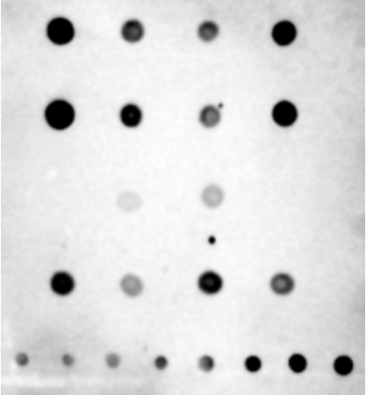
Example membrane image showing IVT samples and the poly(dI) positive control.
8. Notes
Section titled “8. Notes”The run is acceptable when the dsRNA positive control gives a detectable signal, every sample and control position is clearly identifiable, and the compared samples carried equivalent total RNA.
- The J2 antibody recognises dsRNA regions longer than about 40 nucleotides.
- Prepare 5 % milk-TBST fresh on the day.
- Keep the membrane fully covered through every antibody incubation and wash.
- Include a dsRNA positive control in every experiment.
9. References
Section titled “9. References”- Bhaskaran H. Work Instructions for Dot-Blot to Detect Levels of dsRNA. RNA Sciences WI-005, revision 2, effective 22 June 2024.
- Thermo Fisher Scientific. iBright desktop analysis software.