Nicking Insertion Confirmation
The following insertion confirmation strategy is used to confirm the insertion of oligonucleotides in the internal sequence of lambda using Nicking Insertion. This strategy is adapted from a similar strategy used by Gorman and Greene in the following mismatch repair paper Gorman et.al.
In order to re-create this protocol, You must have TS lambda that has not undergone any oligonucleotide insertion, TS lambda with your experimental oligonucleotide inserted, and a negative control generated by inserting an oligonucleotide of the full complement of the insertion site. A previously characterized positive control would also help.
Contents
Section titled “Contents”1. Overview
Section titled “1. Overview”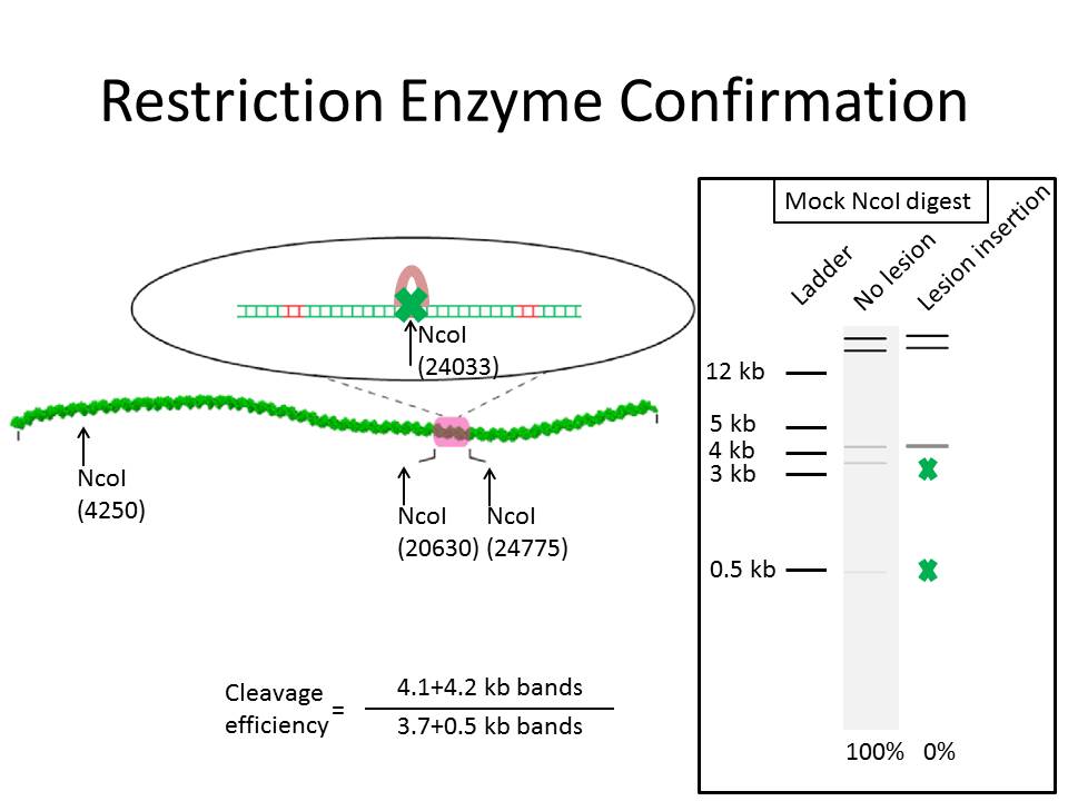
The following figure represents a breakdown of the protocol as well as some hypothetical results. The cartoon on the left represents the restriction enzyme inhibition strategy. Insertion of a mismatch in an NcoI restriction site, indicated by the green X over the loop like structure, will prevent the cleavage of this site by NcoI. This will lead the change in the digestion profile between TS lambda that has not had any oligonucleotides inserted vs. those that have. To quantify the efficiency of the insertion, The band intensity of the inhibited bands are measured relative to the persistent bands. This equation is found below the cartoon. This value is then reported relative to completely digested TS lambda control. The fake gel on the right represents what these results should look like.
2. Oligonucleotide Design
Section titled “2. Oligonucleotide Design”This is the top strand of the insertion site that was cloned into Lamba by Tim Silverstein of the Greene Lab. It was cloned in between them NgoMIV/XbaI sites in the original lysogen.
5’-ATTTAAATAAAGCTCTTCATGCATGCGGCCGCTCTTCCCATGGTGCGATCGCTCTTCGGGATTTAAATA-3’
The bold sections are the recognition sequence of NT.BspQI. The italicized section is the critical NcoI restriction. The italicized and underlined cytosine is the critical amino acid that if interrupted by a single base mismatch or other structural element greatly reduces the efficiency of cleavage.
After clevage with Nt.BspQI, your top strand should look like this
5’-ATTTAAATAAAGCTCTTCA—————————————-GGATTTAAATA-3’
This is the step in which you add your insertion oligonucleotides. Your insertion oligonucleotides should include the sequence you want as well as an interrupt at the critical NCOI cytosine. This can be interrupted in your insert sequence, or with a simple single base pair mismatch. For example, the following mismatch has been tested and works as a good positive control for cleavage inactivation.
5’-CTCTTCATGCATGCGGCCGCTCTTCCTATGGTGCGATCGCTCTTCG-3’
In this insertion oligonucleotide, the cytosine is replaced by a thymine.
3. Restriction digestion
Section titled “3. Restriction digestion”The Digestion is very straight forward. The number of samples you will have will be then number of experimental oligonucleotides you tried to insert +4 (a NEB lambda control, a TS lambda control, a full complement - control, and a single mismatch + control. For each sample, there will be 3 reactions. A non-digested control, a NcoI digestion, and a PvuI-HF digestion. The PvuI-HF digestion is to prove the recombination via the addition of a unique PvuI-HF site into lambda.
Reactions are the following composition:
- 4 ng of DNA (8ul of NEB and control TS lambda, 25ul of inserted TS lambda)
- 2.5ul of NEB #3
- for control reactions, dilute final volume up to 25ul.
NcoI digests use 2.5ul of NcoI and digest for 10min PvuI-HF reactions use 2.5ul of PvuI-HF and digest for 3 hours Controls incubate for 3h with PvuI-HF
Quench the reactions with 1mM EDTA. Load 12.5 ul of the sample into an 0.8% agarose gel. Gels run for either 30 or 45 minutes and are visualized with EtBr.
4. Representative results
Section titled “4. Representative results”The following are a sample of the final product.
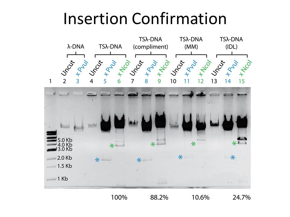
The lane identities are listed above the gel. Blue * highlight the NcoI expected digestion. Green * highlights one of the inhibited NcoI cleavage bands. The second inhibited band is running of the gel.